Special Eye Testing
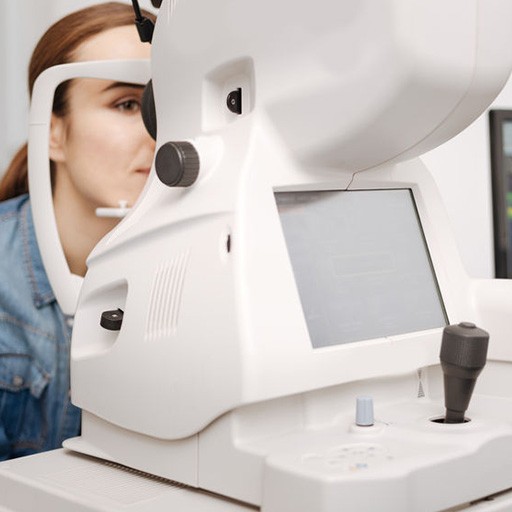
Optical Coherence Tomographer (OCT)
Our OCT helps us better manage glaucoma and diseases of the retina because this technology allows the eye doctor to see the deep tissue layers in the eye. Similar to ultrasound, this diagnostic technique employs light rather than sound waves to achieve higher resolution pictures of the structural layers of the back of the eye. These high-definition images are the only way that they can actually see beneath the surface to the nerve fiber layers where damage occurs. Up until now, eye doctors had to use other tests to indicate damage in this critical area of sight. Common eye diseases such macular degeneration, diabetic retinopathy, and glaucoma are detected early by the OCT when the diseases can be more effectively treated.
Humphrey Visual Field Testing
Visual Field testing can help save vision because it is another test used to diagnose or rule out glaucoma and other neurological disorders that affect vision. This simple, but effective service has saved lives by detecting various medical conditions such as strokes, brain tumors, and other neurological defects.
Visually Evoked Potential (VEP)
The VEP allows the doctor to more accurately diagnose and manage optic and nerve diseases. This advanced technology actually measures the neurological responses of the entire visual pathway, giving a more accurate diagnosis and vision and nerve disorders such as amblyopia and glaucoma.
Retinal Digital Imaging – Fundus Photography
A high-definition digital image of the retinal area helps your eye doctor in Wytheville, Pulaski, Bluefield & Galax, VA diagnose and manage eye diseases in the delicate retinal area. Damage to these delicate structures of the retinal area is often the first sign of systemic diseases such as MS, diabetes and more. The retina is the “window to the body” and routine retinal imaging can help your eye doctor monitor the changes in your eye health from year to year.
B Scan Ultra Sound
The B Scan can save your vision by providing a better look at the back of the eye for signs of retinal detachments and tumors. B scan ultrasound helps your eye doctor with clinical assessment of various eye diseases. The B Scan allows your eye doctor to see structures in the back of the eye that may be obscured by cataracts or blood.
A Scan Ultra Sound
The A Scan ultra sound is a routine test used to determine the length of the eye and is used for calculations of intraocular lens implants prior to cataract surgery.
Specular Microscopy
Specular microscopy is a gentle, imaging technique that allows your eye doctor to diagnosis and monitor diseases of the cornea. These precise images use light to provide data about the number and health of the cells of the cornea.
Our Relationship with local Specialists keeps you seeing your best.
LASIK Vision Correction
Our eye doctors will provide detailed consultations to determine if you are a candidate for laser vision correction. Local surgeons provide the laser correction and our eye doctors handle all post-operative visits and subsequent exams.
Advanced Retinal Care
Wythe Eye Associates offers in house advanced retinal care for diabetic retinopathy, wet macular degeneration, retinal detachment, and other retinal disorders by partnering with Vistar Retinal Specialists.
Cataract Surgery
Wythe Eye Associates is partnered with Vistar to provide advanced cataract surgery and surgeries of the eye. Vistar provides consulatations and evaluations at Wythe Eye Associates to determine candidacy for cataract surgery